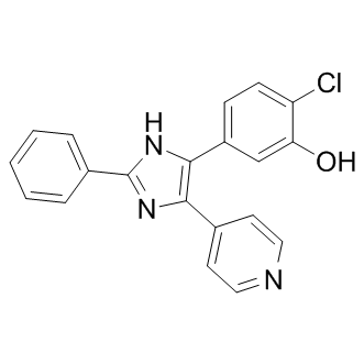
One such example is Selenoprotein S. SelS was first identified in a screen to find genes that were differentially expressed in a diabetic animal model, although it was not yet recognized as a selenoprotein. It was shown to be a glucose-regulated protein, with its expression inversely proportional to circulating glucose and insulin levels. Recently, SelS was identified as one of the most widespread eukaryotic EX 527 selenoReversine proteins based on comparative genomics. It was grouped in a protein family with Selenoprotein K, based on protein localization, domain organization and placement of Sec near the carboxy-terminus. The combination of the prevalence and conservation of SelS suggests that this protein performs an important biological function. The ability of SelS to act as a reductase was demonstrated in vitro, but an enzymatic activity for this protein has not been identified in cells. However, SelS was discovered to play a role in the unfolded protein response. The UPR refers to a group of conserved signaling pathways that are activated in response to the accumulation of unfolded proteins within the ER. The purpose of the UPR is to restore the ability of the ER to process its client proteins, both through the upregulation of molecular chaperones to increase folding capacity and the removal of misfolded proteins to reduce demand. SelS is involved in ERAD as part of a multiprotein complex that removes misfolded proteins from the ER to the cytoplasm for degradation. SelS is also known as Valosin-containing protein Interacting Membrane Protein due to its interaction with VCP in this ERAD complex. The expression of SelS is upregulated under conditions of ER stress, presumably to help increase the capacity of a cell to manage misfolded proteins. The UPR is a crucial cellular pathway as failure to resolve ER stress will cause the cell to undergo apoptosis. Studies in multiple systems have shown that overexpression of SelS has protective effects against ER stress, while knockdown of SelS sensitizes cells to ER stress and apoptosis. Endogenously, this increase in SelS expression is facilitated by the presence of an ER-stress element in its promoter. A naturally occurring point mutation within the ERSE of SelS led to the discovery of a second physiological function. Patients with this mutation were unable to upregulate SelS expression under ER stress conditions. These patients had increased inflammation as determined by plasma levels of  IL-6, IL-1b and TNF-alpha, three acute phase cytokines. This inverse relationship between the expression of SelS and acute phase cytokines suggests that SelS has a role in the negative regulation of inflammation. Furthermore, siRNA knockdown of SelS in macrophage cells led to increased release of IL-6 and TNF-alpha, while treatment of HepG2 cells with cytokines increased SelS expression. This suggests the existence of a regulatory feedback loop to control inflammatory processes. An additional line of evidence linking SelS to inflammation is its direct interaction with serum amyloid A, an acute-phase inflammatory response protein, though the significance of this interaction is unknown. ER stress and inflammation are now known to underlie many human diseases with examples that include diabetes, metabolic syndrome disorders, atherosclerosis, Alzheimer’s Disease, Parkinson’s Disease and non-alcoholic fatty liver disease.
Understanding the molecular mechanisms that contribute to the developmen for selenoproteins are continually being discovered
Leave a reply