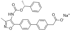
In endothelial cells can also induce angiogenensis in a dose-dependent manner and also RENCA cell proliferation in the present study. Thus activation/ inactivation of LacCer synthase by agonists/antagonists may well regulate angiogenesis in vitro and in vivo. Our BIBW2992 studies and those of others have shown that D-PDMP is non-toxic when given at doses ten times that of the concentration used in the present study. The body weight in this study did not differ in placebo vs. D-PDMP�Ctreated mice. The tumor weight decreased approximately 50% in 3 MPK and 10 MPK fed mice compared to placebo. However, when mice were fed higher amounts of D-PDMP; 25 and 50 MPK, it did not further reduce tumor volume. In a previous study, it was shown that the t1/2 of D-PDMP in mice blood is,50 min. Consequently, it is feasible that  beyond a threshold of 10 MPK, most of this compound is rapidly removed by excretion and therefore further reduction in tumor volume was not observed. Previously, D-PDMP has been used extensively to examine the role of glycosphingolipid and related glycosytransferases in arterial smooth muscle cell proliferation, wound healing, osteoclastogenesis, Nutlin-3 polycystic kidney disease, elasticity, respiratory diseases, glioblastoma research cholesterol efflux, inflammation in vitro and in vivo, shear stress, and A beta secretion in neuroblasotma cells. Although D-PDMP is known to inhibit the activity of UGCG, raise ceramide levels and induce cell death by apoptosis-we could not reproduce these observations in vivo in mice kidney. In agreement with a previous study we also observed that the level of ceramide in kidney in D-PDMP �Ctreated mice was lower. Likewise, an iminosugar, another inhibitor of UGCG also did not raise the level of ceramide in a transgenic mouse model of hyperlipidemia. Moreover, in a recent study, the use of another glucosylceramide synthase inhibitor, Genz122346, in a mouse model of polycystic kidney disease revealed that this compound also inhibits proliferation but does not inhibit apoptosis involving ceramide. This could be due to further catabolism of ceramide as the activity of several hydrolases including ceramide deacylase maybe higher upon treatment with D-PDMP. Also ceramide may be converted to other sphingolipids. These observations attest to the multiple fates of ceramide and multiple pools of ceramide in kidney tissue. Indeed, we observed that the activity of GlcCer glucosidase was increased in D-PDMP�Ctreated mice compared to placebo. This may have contributed to an increase in the level of GlcCer in mice fed D-PDMP. We have previously shown that in cultured human arterial endothelial cells, D-PDMP can inhibit VEGF-induced angiogenesis and this was bypassed by LacCer but not S-1-P. Such observations suggest that LacCer mediated and VEGF-induced angiogenesis is independent of S-1-P-induced angiogenesis. Moreover, use of 1-phenyl-2-palmitoylamino-3-morpholino-1propanol ; a relatively more specific inhibitor of GlcCer synthase compared to D-PDMP to mitigate VEGF induced angiogenesis was bypassed by feeding LacCer in human endothelial cells. In fact VEGF-induced angiogenensis in these cells were mitigated,1.5 fold better by the use of D-PDMP compared to PPMP. Finally, LCS gene ablation by the use of siRNA mitigated VEGF induced angiogenesis in these cells. In the present study, we document that D-PDMP may well inhibit angiogenesis by way of mitigating the expression of p-AKT-1 and mTOR expression in mice kidney.
Collectively our observations imply that the previously reported that L-PDMP which activates LacCer synthase
Leave a reply