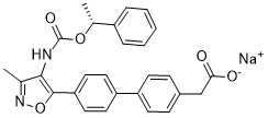
Together, this provides evidence that Metnase could play a role in the pathogenesis and resistance of metastatic breast cancer to Topo IIa inhibiting therapies. Since Metnase enhances Topo IIa-mediated decatenation, and enhances resistance to ICRF-193 and VP-16 in non-malignant human cells, we hypothesized that Metnase might also promote resistance to the anthracyclines and epididophyllotoxins in MDAMB-231 cells. We first investigated whether reducing Metnase would affect ICRF-193-mediated metaphase arrest. MDA-MB-231 cells were treated with ICRF-193, which inhibits Topo IIa after DNA religation, and therefore does not induce DSBs but does inhibit decatenation, allowing for discrimination between DNA damage and metaphase arrest. The increase in cells arrested at metaphase in the presence of ICRF-193 compared to vehicle controls provides a measure of cells arrested due to failure of decatenation. Using atubulin immunoR428 fluorescence microscopy, we determined the fraction of cells in metaphase after exposure to ICRF-193. Cells with reduced Metnase expression showed  a significantly higher percentage of metaphase arrested cells when treated with ICRF193 and cytospun onto slides to retain all cells.After 18 hour treatments with 2 or 10 mM ICRF-193, or 4 hours with 10 mM ICRF-193, cells with reduced Metnase showed 4.9-fold, 2.2-fold, and 2.6-fold increased metaphase arrest, respectively, as compared to vector control and evaluated by student’s t-test. This result suggests that Metnase promotes decatenation in ICRF-193-treated MDA-MB-231 cells, allowing them to proceed through metaphase even in the presence of this Topo IIa specific inhibitor. Prior studies revealed that bladder and lung cancer cells progress through the decatenation checkpoints when Topo IIa is inhibited by high concentrations of ICRF-193. The conclusion from those studies was that these cancer cells failed to arrest because they had inactivated the decatenation checkpoints. While the ability to progress through mitosis even when Topo IIa is inhibited may be a general feature of malignancy, it may be due to the presence of Metnase alone, or Metnase in combination with checkpoint inactivation. Thus, the decatenation checkpoint may be intact in these malignant cells, but Metnase promotes continued Topo IIa function despite the presence of inhibitors, and the decatenation checkpoint is not activated. The Topo IIa inhibitor ICRF-193 does not induce significant DNA damage, and therefore is not relevant in the clinical therapy of breast cancer. To determine whether altering Metnase levels would affect resistance to clinically relevant Topo IIa inhibitors, such as VP-16 and adriamycin, we determined the cytotoxicity of these agents in MDA-MB-231 cell lines that stably under-expressed Metnase using colony formation assays. Decreased Metnase expression increased sensitivity 7.5-fold to VP-16, and 3.5-fold to adriamycin. Together, these results indicate that Metnase expression levels directly correlate with cell survival after exposure to these clinically relevant Topo IIa inhibitors. Adriamycin is an important agent in both adjuvant therapy and in the treatment of metastatic breast adenocarcinoma, so this finding is of relevance for current clinical ICG-001 Wnt/beta-catenin inhibitor regimens. It raises the possibility that treatment efficacy could be improved if the drug was used in combination with a future Metnase inhibitor, or if Metnase levels could be measured and possibly account for variance in responsiveness to adriamycin based chemotherapeutic regimens. Altogether, these results provide further support for the hypothesis that Metnase plays a key role in Topo IIa function. To determine the mechanism for the ability of Metnase to mediate sensitivity to Topo IIa inhibitors, we investigated whether Metnase levels affected the cellular apoptotic response to adriamycin. We exposed MDA-MB-231 cells to adriamycin for 24 hrs and then evaluated annexin-V/FITC fluorescence by flow cytometry.
shRNA down-regulation of Metnase levels markedly sensitized to adriamycininduced immunoprecipitate with Topo IIa
Leave a reply